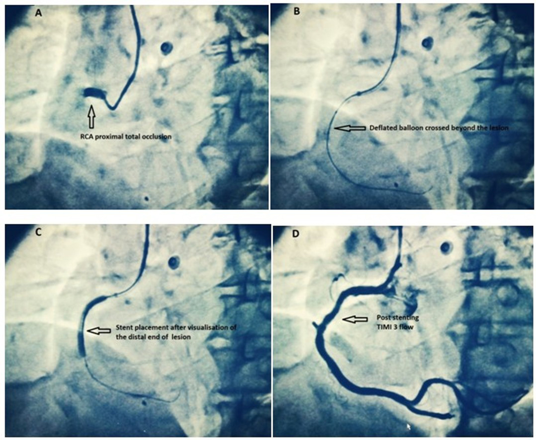
In order to quantitatively characterize the kinetics of dye entering the myocardium using the angiogram, digital subtraction angiography (DSA) was developed. Those patients with both TIMI grade 3 flow and TIMI myocardial perfusion grade 3 flow had a mortality of 0.7% while those patients with both TIMI grade 0/1 and TIMI myocardial perfusion grade 0/1 flow had a mortality of 10.9% (4). The TIMI flow grades and the TIMI myocardial perfusion grades can be combined to identify a group of patients at very low risk and alternatively very high risk for mortality. In contrast, those patients with both TIMI grade 3 flow in the epicardial artery and TIMI myocardial perfusion grade 3 have a mortality under 1% (4). Despite achieving epicardial patency with normal TIMI grade 3 flow, those patients whose microvasculature fails to open (TIMI myocardial perfusion grade 0/1) have a persistently elevated mortality of 5.4%. The TMPG permits risk stratification even within epicardial TIMI grade 3 flow. The TMPG has been shown to be a multivariate predictor of mortality in acute MI (Fig. There is either minimal or no ground glass appearance (blush) or opacification of the myocardium in the distribution of the culprit artery indicating lack of tissue level perfusion. TMPG 0: Dye fails to enter the microvasculature. There is the ground glass appearance (blush) or opacification of the myocardium in the distribution of the culprit lesion that fails to clear from the microvasculature, and dye staining is present on the next injection (approx 30 s between injections). TMPG 1: Dye slowly enters but fails to exit the microvasculature. There is the ground glass appearance (blush) or opacification of the myocardium in the distribution of the culprit lesion that is strongly persistent at the end of the washout phase (i.e., dye is strongly persistent after three cardiac cycles of the washout phase and either does not or only minimally diminishes in intensity during washout).

TMPG 2: Delayed entry and exit of dye from the microvasculature. Blush that is of only mild intensity throughout the washout phase, but fades minimally, is also classified as grade 3. There is the ground glass appearance (blush) or opacification of the myocardium in the distribution of the culprit lesion that clears normally, and is either gone or only mildly/moderately persistent at the end of the washout phase (i.e., dye is gone or is mildly/moderately persistent after 3 cardiac cycles of the washout phase and noticeably diminishes in intensity during the washout phase), similar to that in an uninvolved artery. TMPG 3: Normal entry and exit of dye from the microvasculature. This led to the recent development of a new angiographic measure of tissue level perfusion, the TIMI myocardial perfusion grade (TMPG) (4), as follows: It has recently become apparent that epicardial flow does not necessarily imply tissue level or microvascular perfusion (21,22).


 0 kommentar(er)
